By Sayer Ji
Contributing writer for Wake Up World
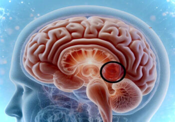
A recent analysis found over 60% of people have calcification of the pineal gland, which produces the hormone melatonin regulating sleep-wake cycles. Calcification associates with melatonin decline, health issues.
[pro_ad_display_adzone id=”110028″]
The Calcifying Effects of Fluoride on the Pineal Gland
The pineal gland has been referred to as the “seat of the soul” for hundreds of years due to its light-sensitive nature and production of the spiritual hormone melatonin[1]. But could fluoride, a ubiquitous modern-day toxicant, be calcifying this gland and literally turning our spiritual center to stone?[2] Emerging research suggests the answer is yes.
In a 2001 study on fluoride distribution, pineal gland fluoride levels were measured in aged cadavers. Researchers found that:
“There was a positive correlation between pineal F[luoride] and pineal Ca[lcium] (r = 0.73, p<0.02) but no correlation between pineal F and bone F. By old age, the pineal gland has readily accumulated F and its F/Ca ratio is higher than bone.“[3]
They concluded that over time through regular fluoride exposure, “the pineal accumulates fluoride and that this accumulation may be implicated in the pathogenesis of pineal calcification.”[3] Given the essential role of the pineal gland and melatonin in regulating wake/sleep patterns and spiritual health[4], these findings have profound implications.
The pineal gland produces the vital spiritual hormone melatonin, which regulates healthy sleep-wake cycles[5]. Studies reveal calcification is associated with decreased nighttime melatonin production[6]:
“Calcification of the pineal gland is shown to be closely related to defective melatonin secretion, which enhances oxidative stress.“[7]
Decreased melatonin is subsequently linked with various sleep cycle disturbances including daytime tiredness:
.png)
“[Calcification] is associated with decreased REM sleep percentage, decreased total sleep time, poorer sleep efficiency, greater sleep disturbance and greater daytime tiredness.“[8]
Animal studies also demonstrate that pineal gland removal results in loss of response to the anti-depressant Prozac[9], suggesting disturbing effects on mood regulation as well:
“The mechanism behind mood stabilization and antidepressant effect of fluoxetine may be abolished in pinealectomized rats due to loss of circadian rhythm of plasma melatonin levels.”[10]
Pineal gland fluoride can also contribute to free radical damage and inflammation, thereby inflaming conditions like depression and other disorders:
“Fluoride leads to generation of free radicals and enhanced lipid peroxidation in animals and humans through oxidative mechanisms.“[7]
Meanwhile melatonin has direct antioxidant effects offering protection. But with enough accumulated fluoride, the pineal gland’s essential detoxifying and regulatory roles are hindered, which undoubtedly impacts the soul.
So how exactly does fluoride infiltrate the seat of the soul? The pineal gland protrudes from the center of the brain and uniquely lacks the blood-brain barrier protecting most of the brain from bloodstream toxins like fluoride[2]. Pineal gland tissue also contains high levels of hydroxyapatite, which attracts circulating fluoride like a magnet and causes it to accumulate over time[2,11]:
“Fluoride has a great affinity toward hydroxyapatite crystals, so the pineal (which calcifies early in life) accumulates fluoride over time.”[12]
Animal studies reveal eliminating fluoride exposure for as little as 5 weeks stimulates significant pineal gland repair and reverses accumulated damage, underscoring the importance of reducing sources of toxic fluoride[13]. Similar healing would likely occur in human glands as well.
Beyond avoiding sources like fluoridated water, fluoride dental products, non-stick pans, and certain drugs, increasing intake of iodine, magnesium, selenium, vitamin K2, and antioxidants like melatonin helps mitigate pineal gland accumulation and promote healthy de-calcification[14]. With wise prevention and periodic detox from this ubiquitous toxicant, we can sustain the seat of the soul for future spiritual connection.
Recent Systematic Review Finds High Global Prevalence of Pineal Gland Calcification
.png)
A 2023 systematic review and meta-analysis published in Systematic Reviews aimed to assess the pooled global prevalence of pineal gland calcification (Pineal Res. 2023; 12:32). The authors conducted a comprehensive literature search and analysis of population-based studies reporting prevalence rates.
From 30 studies screened, 8 cross-sectional studies representing over 5,500 patients were included in the final quantitative analysis. The pooled prevalence of pineal gland calcification was found to be 61.65% (95% CI: 52.81-70.49%) with high heterogeneity between studies (I2 = 97.7%).
Reported prevalence rates ranged widely from 26.88% to 76.7% between countries. Qualitative review revealed pineal calcification consistently increased with age, was more common in males versus females, and was higher among white ethnicity.
While specific causes remain unclear, contributors likely include genetic, environmental, lifestyle and neurodegenerative factors. Potential mechanisms span accumulating hydroxyapatite deposits or cellular debris within the gland’s unique physiology alongside calcifying processes seen in bone and vasculature.
Given the pineal gland’s role regulating sleep-wake cycles and other rhythms via spiritual hormone melatonin, these high rates of calcification have profound clinical implications. Pineal concretions associate dose-dependently with melatonin decline, circadian dysfunction, sleep disturbances and psychiatric symptoms.
[pro_ad_display_adzone id=”110030″]
Future studies must clarify specific causes and impacts of rising pineal gland calcium accrual both globally and in vulnerable subpopulations. Since pineal physiology remains incompletely understood, the prevalence, predictors and clinical sequela of pineal calcification deserve fuller investigation.
In summary, this systematic analysis established significantly high worldwide prevalence of pineal gland calcification using the largest dataset to date. These findings highlight an urgent need to elucidate the drivers and consequences of pineal concretions given the essential spiritual and homeostatic functions governed by this unique neuroendocrine organ.
Decalcifying Soft Tissue and Supporting Pineal Health
Since calcification may contribute to conditions like Alzheimer’s disease and pineal gland dysfunction, evidence-based natural interventions to remove excess calcium deposits deserve exploration. The research database GreenMedInfo.com contains research on over 95 natural substances studied to reduce soft tissue calcification, with top evidence for magnesium, garlic and vitamin K2.[16]
.png)
Reducing intake of inorganic calcium supplements could also mitigate risk, as excessive supplementation associates with pineal calcification and lesions in the brain.[17] The use of inorganic calcium supplements, even at low doses, correlates with greater volume brain lesions compared to non-users.[18]
Considering the pineal gland’s melatonin-producing effects on sleep-wake and antioxidant activity, supporting its structural health aligns with evidence-based natural healing. Beyond moderate sun exposure for optimal melatonin, dietary sources like walnuts, oranges and kiwi support healthy circadian rhythms and sleep quality.[19] Herbs including lavender, passionflower and valerian root as well as supplemental melatonin also promote sound sleep and brain health through the aging process.[20]
References:
[1] Macchi & Bruce (2004). Human pineal physiology-seminal review. Front Neuroendocrinol, 25(3-4), 177-195. https://pubmed.ncbi.nlm.nih.gov/15381018/
[2] Chlubek & Sikora (2020). Fluoride and the pineal gland-seminal paper. Appl Sci, 10(8), 2885. https://www.mdpi.com/2076-3417/10/8/2885
[3] Luke (2001). Fluoride deposition in aged pineal gland-original study. Caries Res, 35, 125-128. https://pubmed.ncbi.nlm.nih.gov/11485891
[4] Tan et al. (2018). Pineal gland aging and melatonin loss-review. Mol, 23(2), 301. https://www.mdpi.com/1420-3049/23/2/301
[5] Zisapel (2018). Melatonin and sleep-authoritative review. Br J Pharmacol, 175(16), 3190-3199. https://pubmed.ncbi.nlm.nih.gov/29624363/
[6] Kunz et al. (1999). Decreased melatonin and pineal gland calcification. Neuropsychopharm, 21(6), 765-72. https://pubmed.ncbi.nlm.nih.gov/10481836/
[7] Shivashankara (2000). Fluoride toxicity and oxidative stress. Fluoride, 33(1), 17-22.
[8] Mahlberg (2009). Pineal gland calcification and sleep measures. Sleep Med, 10(4), 439-445. https://pubmed.ncbi.nlm.nih.gov/19249556/
[9] Mørk et al. (2004) Pinealectomy alters anti-depressant response. Psychopharm, 171(4), 408-15. https://pubmed.ncbi.nlm.nih.gov/14534750/
[10] Zhang (2015). Fluoxetine mood effects via melatonin. Neuroreport, 26(8), 463-9. https://pubmed.ncbi.nlm.nih.gov/25893300/
[11] Tharnpanich (2016). Association of pineal gland fluoride and calcium. Fluoride, 49(4 pt2), 472-484. https://pubmed.ncbi.nlm.nih.gov/27695626/
[12] Ge et al. (2011). Fluoride and intelligence in children-seminal review. Fluoride, 44(4), 190-194.
[13] Mrvelj & Womble (2020). Fluoride elimination stimulates pineal growth-original study. Biol Trace Elem Res, 197(1), 175-183. https://pubmed.ncbi.nlm.nih.gov/31094211/
[14] Spittle (2008). Fluoride and the pineal gland-review. Fluoride, 41(2), 103-104. https://fluoridealert.org/wp-content/uploads/spittle-2008.pdf
[15] Belay DG, Worku MG. Prevalence of pineal gland calcification: systematic review and meta-analysis. Syst Rev. 2023 Mar 6;12(1):32. doi: 10.1186/s13643-023-02205-5. PMID: 36879256; PMCID: PMC9987140.
[16] GreenMedInfo.com. Soft Tissue Calcification (95 natural agents studied). www.greenmedinfo.com/disease/soft-tissue-calcification
[17] Ji, S. Taking Calcium Supplements Causes Brain Lesions. GreenMedInfo Article. www.greenmedinfo.com/blog/taking-calcium-supplements-causes-brain-lesions
[18] Payne, M. E., Anderson, J. J. B., & Steffens, D. C. (2014). Calcium and Vitamin D Intakes May Be Positively Associated With Brain Lesions in Depressed and Nondepressed Elders. Nutrition Research, 34(3), 285-295. https://doi.org/10.1016/j.nutres.2014.01.007
[19] GreenMedInfo.com. Natural Agents with Circadian Rhythm Modulating Activity. www.greenmedinfo.com/disease/circadian-rhythm-disorder/natural-agents-with-circadian-rhythm-modulating-
[20] GreenMedInfo.com. Natural Agents with Sedative Properties. www.greenmedinfo.com/disease/insomnia/natural-agents-with-sedating-properties
About the author:
Sayer Ji is the founder of Greenmedinfo.com, a reviewer at the International Journal of Human Nutrition and Functional Medicine, Co-founder and CEO of Systome Biomed, Vice Chairman of the Board of the National Health Federation, and Steering Committee Member of the Global Non-GMO Foundation.
© 2020 GreenMedInfo LLC. This work is reproduced and distributed with the permission of GreenMedInfo LLC. Want to learn more from GreenMedInfo? Sign up for their newsletter here.
[pro_ad_display_adzone id=”110027″]


